Digital Vascular Imaging (DVI)
DVI is an experimental digital imaging method designed to improve visualisation of haemoglobin-containing vascular structures in photographs of the ocular surface. Its clinical use cases are still emerging, and the processed image should be interpreted as an adjunct to conventional examination rather than as angiography or a stand-alone diagnostic test.
How to do the test
- Use a well-focused, evenly illuminated image of the conjunctiva, limbus or scleral region of interest.
- Avoid overexposure, marked reflections and motion blur, as these may interfere with vascular extraction.
- Position the region to be assessed clearly within the imaging field and acquire the image using a consistent working distance and illumination whenever serial comparison is intended.
- Capture the image using the Bluetooth switch or by pressing any key on the keyboard.
- Review the colour image together with the DVI-processed image. Interpretation should always be made in the context of the clinical examination.
- For follow-up studies, try to reproduce gaze direction, magnification, illumination and the area imaged as closely as possible.
What DVI shows
DVI uses colour information within a digital image to preferentially display vascular structures related to haemoglobin absorption. It does not require an injected dye. The resulting image may make vascular patterns more conspicuous than they are in the original colour photograph.
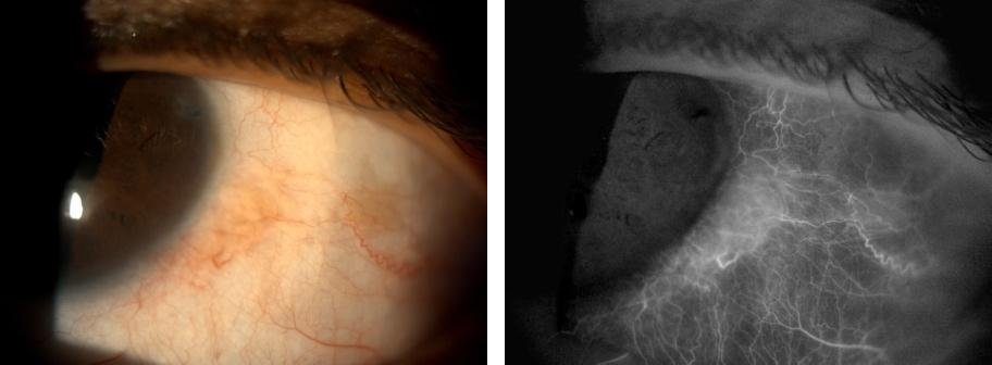
Ocular surface overview
DVI can be used as a documentation aid when comparing the visible-light appearance of the ocular surface with the processed vascular image. This may be useful for reviewing vascular distribution, focal changes, and regional differences over time.
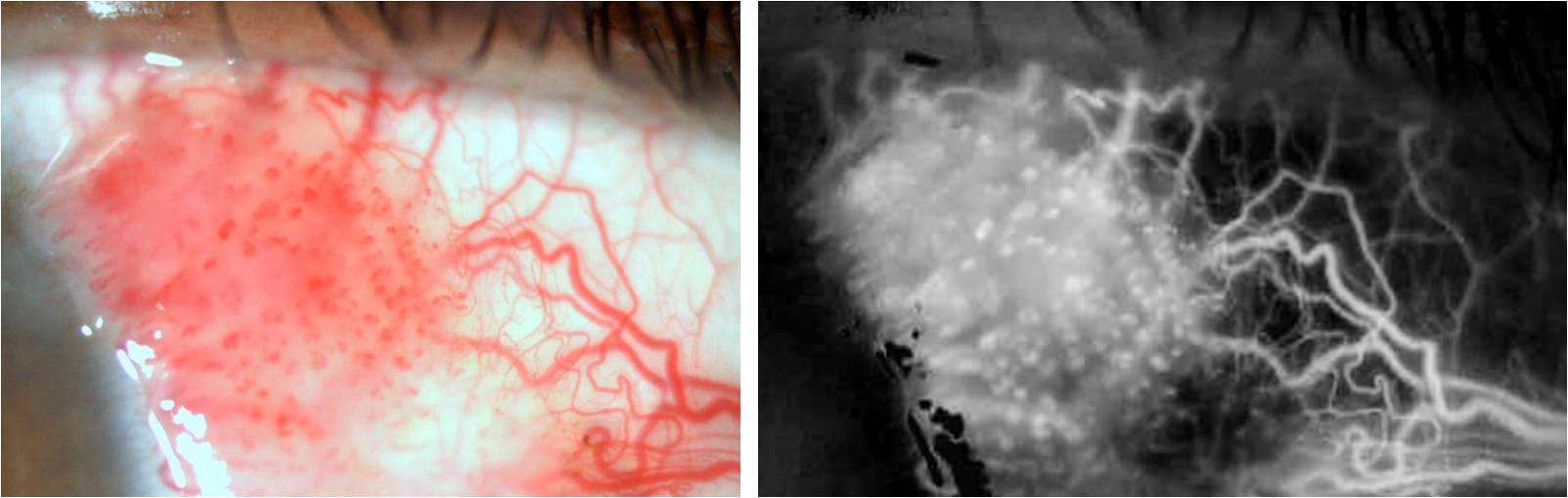
OSSN vascularisation
Ocular surface squamous neoplasia may show prominent intrinsic and feeder vessels. Serial DVI images may be useful for observing changes in lesion-associated vascularisation during treatment and follow-up. Changes in vascular patterns during therapy have also been demonstrated with other vascular imaging techniques such as OCT angiography.1,2
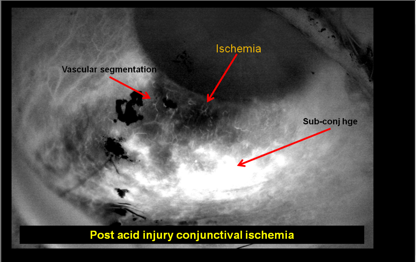
Chemical injury and limbal ischaemia
Limbal ischaemia is an important component of the assessment of severe ocular surface chemical injury and has prognostic significance.3,4 DVI may provide an additional way of documenting the vascular pattern around areas of suspected limbal or conjunctival ischaemia, particularly for serial comparison. This remains an emerging use case and should not replace clinical grading.
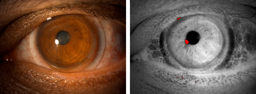
Scleral-lens related blanching
Scleral lenses can produce local conjunctival or limbal vascular compression when the landing zone is poorly aligned; clinically this appears as blanching or interruption of visible vessels.5,6 DVI may help document the distribution of such vascular compression and compare the ocular surface before, during or after lens wear.
Conjunctival vascular patterns
DVI may improve appreciation of vessel density, calibre, branching and regional distribution. Potential applications include documentation of conjunctival hyperaemia, focal vascular changes and longitudinal comparison. Quantitative interpretation requires further validation and standardised acquisition.
Ocular surface tumours
Tumour-associated vascularity can change during therapy. In OSSN, vascular imaging may therefore complement slit-lamp photography when documenting response. DVI does not establish tumour diagnosis, depth or histology, and should not replace clinical examination, anterior-segment OCT or biopsy when these are indicated.
Ischaemia and vascular compression
Areas with reduced visible vascularity can occur after severe chemical injury or from mechanical compression. DVI may make spatial differences in vascular appearance easier to record. Because a processed photograph cannot by itself prove tissue perfusion, the term ischaemia should continue to be based on the complete clinical context.
Research potential
The most promising future direction is likely to be quantitative analysis rather than image appearance alone. Possible measurements include vascular area fraction, vessel length, calibre, tortuosity, branching patterns and regional change over time. Repeatability, calibration and correlation with established clinical measures will be necessary before such parameters can be used diagnostically.
Clinical perspective: DVI is best regarded at present as an adjunctive vascular visualisation and documentation tool. Its potential applications in conjunctival, limbal and scleral disease are evolving.
Selected references
- Liu Z, et al. Optical coherence tomography angiography for visualisation and quantification of vascular networks in ocular surface squamous neoplasia. 2020.
- Theotoka D, et al. OCT angiography assessment of vascular changes in OSSN during topical therapy. The Ocular Surface. 2022.
- Kam KW, et al. Limbal ischaemia: reliability of clinical assessment and implications in ocular burns. 2019.
- Dua HS, et al. Chemical eye injury: pathophysiology, assessment and management. 2020.
- Clinical literature on scleral-lens landing-zone compression and conjunctival vascular blanching.
- Clinical outcomes literature defining scleral-lens limbal compression by disruption of blood flow or scleral blanching.